Hello, in this particular article you will provide several interesting pictures of dimeric structure of pseudokinase rnase l bound. We found many exciting and extraordinary dimeric structure of pseudokinase rnase l bound pictures that can be tips, input and information intended for you. In addition to be able to the dimeric structure of pseudokinase rnase l bound main picture, we also collect some other related images. Find typically the latest and best dimeric structure of pseudokinase rnase l bound images here that many of us get selected from plenty of other images.
 Dimeric Structure of Pseudokinase RNase L Bound to 2-5A Reveals a Basis We all hope you can get actually looking for concerning dimeric structure of pseudokinase rnase l bound here. There is usually a large selection involving interesting image ideas that will can provide information in order to you. You can get the pictures here regarding free and save these people to be used because reference material or employed as collection images with regard to personal use. Our imaginative team provides large dimensions images with high image resolution or HD.
 Dimeric Structure of Pseudokinase RNase L Bound to 2-5A Reveals a Basis dimeric structure of pseudokinase rnase l bound - To discover the image more plainly in this article, you are able to click on the preferred image to look at the photo in its original sizing or in full. A person can also see the dimeric structure of pseudokinase rnase l bound image gallery that we all get prepared to locate the image you are interested in.
 Dimeric Structure of Pseudokinase RNase L Bound to 2-5A Reveals a Basis We all provide many pictures associated with dimeric structure of pseudokinase rnase l bound because our site is targeted on articles or articles relevant to dimeric structure of pseudokinase rnase l bound. Please check out our latest article upon the side if a person don't get the dimeric structure of pseudokinase rnase l bound picture you are looking regarding. There are various keywords related in order to and relevant to dimeric structure of pseudokinase rnase l bound below that you can surf our main page or even homepage.
 Dimeric Structure of Pseudokinase RNase L Bound to 2-5A Reveals a Basis Hopefully you discover the image you happen to be looking for and all of us hope you want the dimeric structure of pseudokinase rnase l bound images which can be here, therefore that maybe they may be a great inspiration or ideas throughout the future.
 Dimeric Structure of Pseudokinase RNase L Bound to 2-5A Reveals a Basis All dimeric structure of pseudokinase rnase l bound images that we provide in this article are usually sourced from the net, so if you get images with copyright concerns, please send your record on the contact webpage. Likewise with problematic or perhaps damaged image links or perhaps images that don't seem, then you could report this also. We certainly have provided a type for you to fill in.
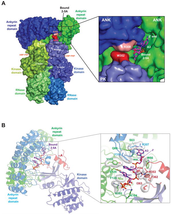
 Structure of the Pseudokinase Domain of RNase L (A) Ribbons view of the The pictures related to be able to dimeric structure of pseudokinase rnase l bound in the following paragraphs, hopefully they will can be useful and will increase your knowledge. Appreciate you for making the effort to be able to visit our website and even read our articles. Cya ~.
 Dimeric Structure of Pseudokinase RNase L Bound to 2-5A Reveals a Basis Dimeric Structure of Pseudokinase RNase L Bound to 2-5A Reveals a Basis
 Dimeric Structure of Pseudokinase RNase L Bound to 2-5A Reveals a Basis Dimeric Structure of Pseudokinase RNase L Bound to 2-5A Reveals a Basis
 Dimeric Structure of Pseudokinase RNase L Bound to 2-5A Reveals a Basis Dimeric Structure of Pseudokinase RNase L Bound to 2-5A Reveals a Basis
 Dimeric Structure of Pseudokinase RNase L Bound to 2-5A Reveals a Basis Dimeric Structure of Pseudokinase RNase L Bound to 2-5A Reveals a Basis
 Dimeric Structure of Pseudokinase RNase L Bound to 2-5A Reveals a Basis Dimeric Structure of Pseudokinase RNase L Bound to 2-5A Reveals a Basis
 Structure of the Pseudokinase Domain of RNase L (A) Ribbons view of the Structure of the Pseudokinase Domain of RNase L (A) Ribbons view of the
 Dimeric Structure of Pseudokinase RNase L Bound to 2-5A Reveals a Basis Dimeric Structure of Pseudokinase RNase L Bound to 2-5A Reveals a Basis
 Table 1 from Dimeric Structure of Pseudokinase RNase L Bound to 2-5A Table 1 from Dimeric Structure of Pseudokinase RNase L Bound to 2-5A
 (PDF) Dimeric Structure of Pseudokinase RNase L Bound to 2-5A Reveals a (PDF) Dimeric Structure of Pseudokinase RNase L Bound to 2-5A Reveals a
 Dimeric Structure of Pseudokinase RNase L Bound to 2-5A Reveals a Basis Dimeric Structure of Pseudokinase RNase L Bound to 2-5A Reveals a Basis
 Dimeric Structure of the Pseudokinase IRAK3 Suggests an Allosteric Dimeric Structure of the Pseudokinase IRAK3 Suggests an Allosteric
 Figure 1 from Structure of the pseudokinase-kinase domains from protein Figure 1 from Structure of the pseudokinase-kinase domains from protein
 Dimeric Structure of the Pseudokinase IRAK3 Suggests an Allosteric Dimeric Structure of the Pseudokinase IRAK3 Suggests an Allosteric
 image/jpeg image/jpeg
 Figure 1 from New insights into the structure and function of the Figure 1 from New insights into the structure and function of the
 Dimeric Structure of the Pseudokinase IRAK3 Suggests an Allosteric Dimeric Structure of the Pseudokinase IRAK3 Suggests an Allosteric
 Protein Kinases: What is the point of pseudokinases? | eLife Protein Kinases: What is the point of pseudokinases? | eLife
 Structure of the pseudokinase-kinase domains from protein kinase TYK2 Structure of the pseudokinase-kinase domains from protein kinase TYK2
 Dimeric Structure of the Pseudokinase IRAK3 Suggests an Allosteric Dimeric Structure of the Pseudokinase IRAK3 Suggests an Allosteric
 Structures of the two dimeric conformers of RNase A (A) 'Opening' of Structures of the two dimeric conformers of RNase A (A) 'Opening' of
 Structure of the Jak1 pseudokinase domain (a) The domain structure of Structure of the Jak1 pseudokinase domain (a) The domain structure of
 The two quaternary structures of BS-RNase Both dimeric structures (one The two quaternary structures of BS-RNase Both dimeric structures (one
 Superposition of the crystal structures of MLKL pseudokinase bound to Superposition of the crystal structures of MLKL pseudokinase bound to
 RNase E recognizes the enolase dimer through a microdomain (a) The RNase E recognizes the enolase dimer through a microdomain (a) The

